


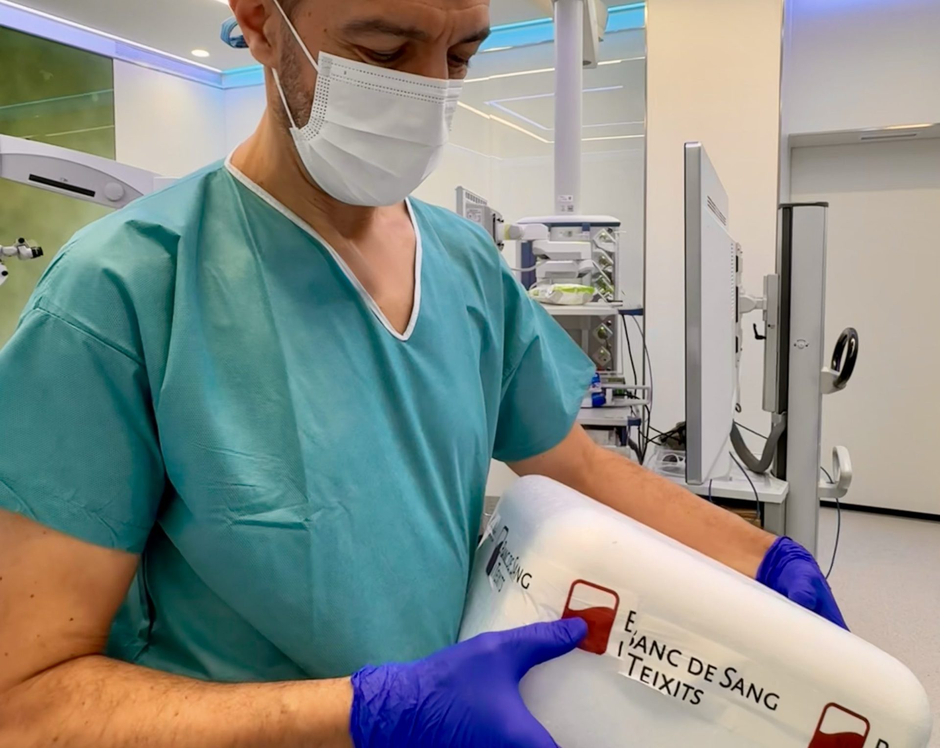
Endothelial cornea transplant: not all cornea transplants are the same
Many people still believe all cornea transplants are the same. They are not. Endothelial keratoplasty replaces only the damaged inner layer, improving recovery—especially in conditions like Fuchs’ dystrophy. As Dr. Josep Torras explains, choosing the right technique is key to both outcomes and patient quality of life.
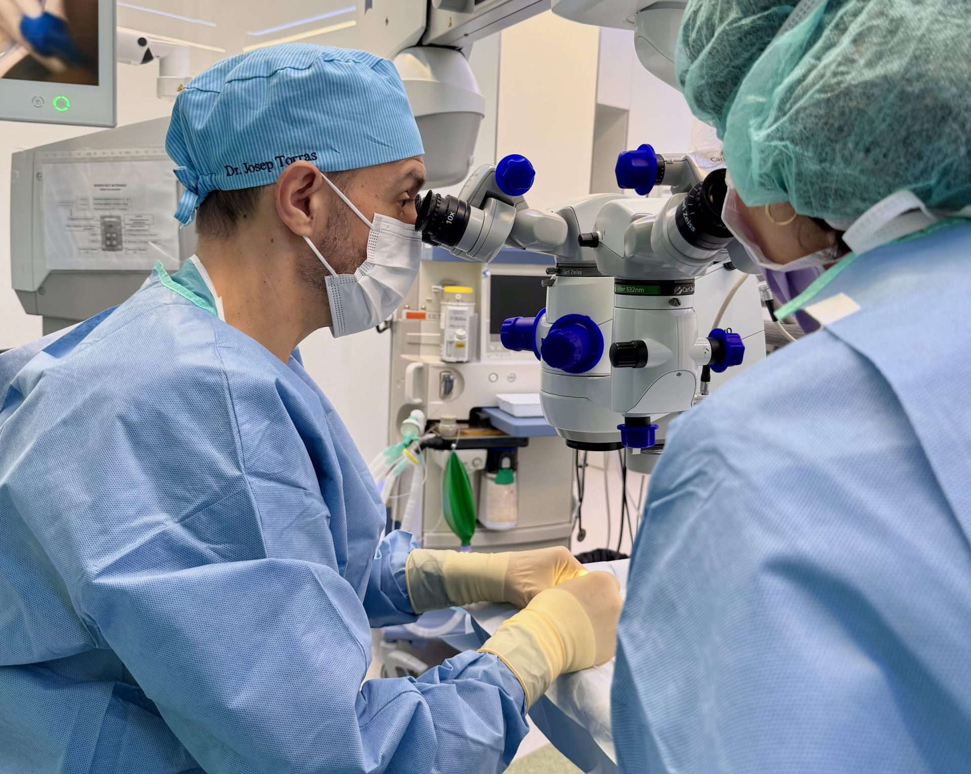
Eye surgery: why choosing the right specialist matters
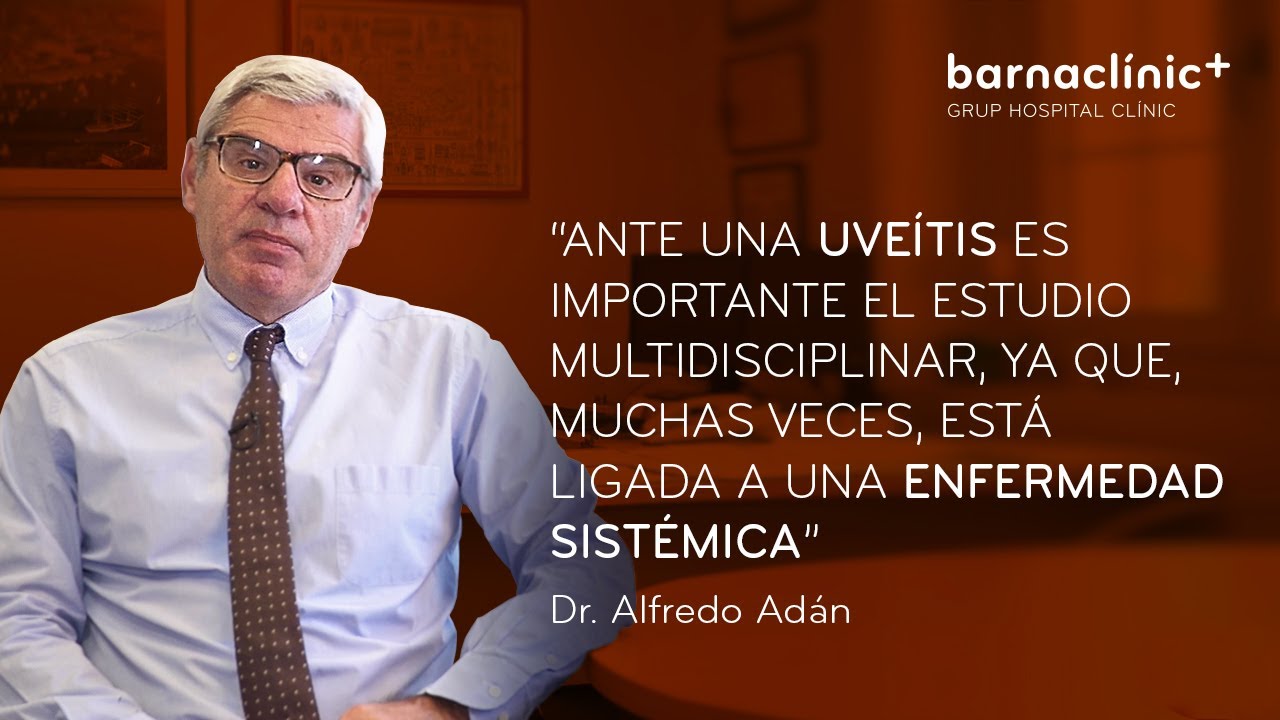
Ophthalmic surgery has advanced so much that it may now seem quick and simple. But behind every procedure there are key decisions that can determine the outcome… and your vision for life. “Thinking that any eye surgery is simple and can be performed by anyone is a mistake,” warns Alfredo Adán.
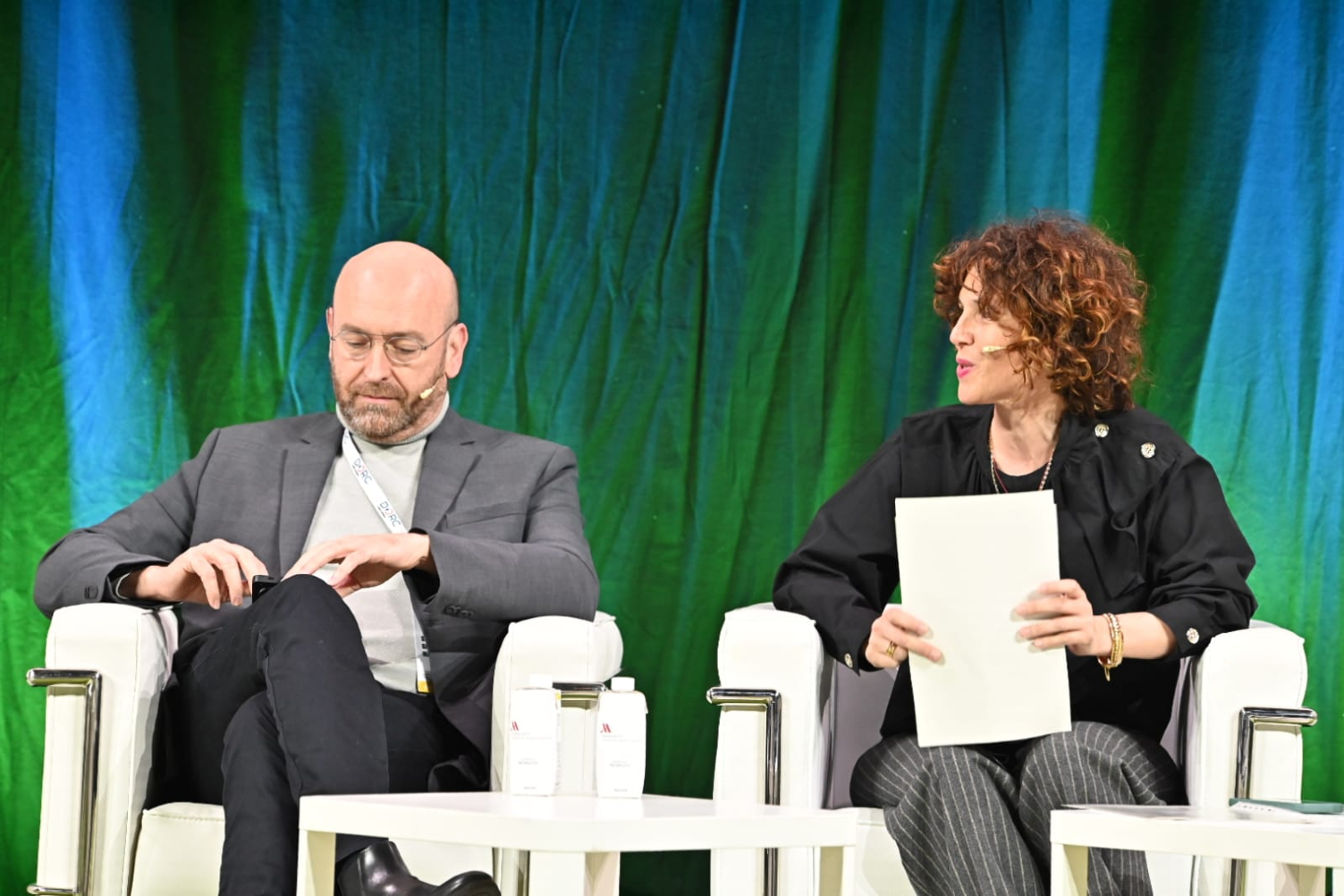
Who would you trust your vision to: someone who applies evidence… or someone who leads it?
Entrusting your vision to those who lead scientific evidence brings a level of reassurance that is hard to explain. Dr. Anna Sala-Puigdollers, retina specialist at Visionclinic, took part in the 29th Congress of the Spanish Society of Retina and Vitreous (SERV), one of the most important scientific meetings in ophthalmology in Spain.


