OCT is an imaging diagnostic technique that, in recent years, has become essential in ophthalmology clinics thanks to its countless advantages. It is simple, non-invasive and facilitates the control and monitoring of any retinal disease. If you suffer from glaucoma or uveitis, surely you have heard of it. If not, Dr Alfredo Adán, director of visiõnclinic+ explains you.
What is an OCT or Optical Coherence Tomography?
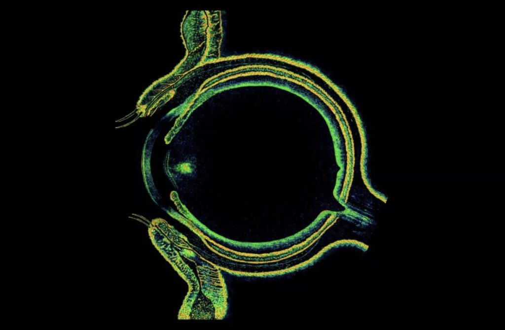
Optical Coherence Tomography or OCT is a simple and non-invasive diagnostic imaging technique that is used for detailed exploration of parts of the retina, specifically the macula, and the optic nerve.

Dr Alfredo Adán
"With micrometric precision, OCT allows us to visualize different ocular structures in 3D, performing an automatic "scanner" that provides us with highly relevant information for the detection and monitoring of many eye diseases"
What are the advantages of OCT?
Its main advantage is that it is a non-invasive technology and with a much higher resolution than other techniques used in medicine that also make transversal optical “slices” over the tissues (such as CT or MRI). For this, the operation of the OCT is based on the emission of infrared light that, when reflected on the ocular structures, shows us the layers or sections of different parts of the eye, detecting very subtle alterations, practically on a cellular scale.
Are there different types of OCT?
Yes, depending on the structures on which it is focused, we can distinguish two types of OCT:
· Posterior pole Optical Coherence Tomography: visualization of the retina and, with the latest models, even of the layer behind it, the choroids.
· Anterior pole Optical Coherence Tomography: visualization of the cornea, the anterior chamber of the eye, the iris and the crystalline lens. It can be done with posterior OCT equipment to which certain software or lenses are adapted, or through new specific equipment for anterior OCT.
How is an OCT performed?
It is a very comfortable and fast test, it lasts approximately between 1 and 3 minutes and is painless, since it does not involve contact with the eye and avoids having to use anesthetic drops. We can perform it without dilating the eye and we obtain the results immediately to complete your diagnosis. We carry it out in our Vía Augusta office, equipped with the latest diagnostic technology.

Dr Marta Pazos
"We have published a study on the efficacy of OCT-based glaucoma diagnostic calculators, making them a key tool in future clinical practice"
What is an OCT used for?
At visiõnclinic we investigate the different uses of this test and its effectiveness in the diagnosis of retinal diseases such as uveitis and glaucoma. In this sense, Alfredo Adán has published several scientific articles that demonstrate its usefulness. Moreover, our glaucoma team, with Marta Pazos, Elena Millá and Néstor Ventura, have just published a study on the efficacy of OCT-based glaucoma diagnostic calculators: “We believe that these tools play a key role in practical clinic”, explains Marta Pazos.